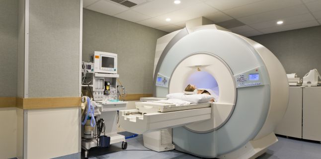
All you need to know about Nuclear Magnetic Resonance (MRI)

Besides the many indications, there are also several categories of people who can not benefit from this investigation. People with a history of allergies to various contrast agents, especially those based on iodine, are a relative contraindication for examination, requiring special measures. Another category that can not benefit from this exploration are pregnant women due to radiation that can be harmful to the fetus. Patients with various metallic implants such as pacemakers, hip prostheses, metal rods, heart valves with metallic structure, or any other metal attached to the body at contact with the magnetic field of the magnetic resonance device may become a potential hazard. Patients who have intrauterine devices such as sterile from various metals should report a doctor prior to the investigation because some do not allow the investigation. Another category of patients who can not do this is the agitated, the frequent convulsions and the patients with renal pathology because they can not receive the contrast substance.
Cranio-cerebral pathology uses this investigation most often, being almost essential for diagnosing. It allows visualization of vascular vessels by angio-NMR and cerebral substance without and with the contrast substance in which causes brain damage: arterio-venous malformations, arterial aneurysms, vasculitis, primary or secondary tumors, post-traumatic lesions, inflammatory processes . MRI spectroscopy provides information on the structure of organic compounds, so magnetic resonance spectroscopy is recommended for patients with various brain disorders such as malignant tumors, benign tumors such as neurinomas, astrocytoma or inflammatory lesion in the brain, metabolic diseases, autoimmune diseases, etc. . This investigation provides a series of information besides scanning the MRI of the brain or spine for the size of the tumor and the chemical structure within the examined tumor.
Therefore, after the investigation and after the radiologist's interpretation, specially colored maps of different colors are created, which accurately show the fixed localization of excessive concentrations of metabolites, which gives very important clues for establishing the correct treatment. with contrast, provides clues about the location of brain lesions, their number, and size, to correctly differentiate between benign and malignant lesions. In the case of benign tumors, a balance is made before and after treatment, and the effectiveness of the treatment is highlighted. This analytical expertise is also useful in distinguishing the degenerative metabolic processes that occur in neurodegenerative disorders such as: vascular dementia, neurosis, etc. .
By monitoring the evolution of lactate in the affected brain area, various types of ischemic or haemorrhagic accidents can be evaluated, as well as hypoxic encephalopathy. MRI tractography investigates the neural tracts of white matter and links with cortical and subcortical regions. This type of MRI investigation evaluates the gradual maturation of the brain over the years. Displaying 3D maps of nerve pathways in white brain is a predictive factor in identifying neurological problems. So transformed into images, tracts will be studied by specialists for accurate and fast diagnosis.
Thus, with the help of Tractography, early detection and monitoring of several neurological conditions such as: stroke (stroke), recent or past trauma, dementia, schizophrenia, but also vascular malformations either clinically inherited or acquired. It can also detect alterations in nerve demyelination, being used in the early diagnosis of demyelinating disorders of nerve fibers and helping the neurosurgeon to establish the preoperative plan of neurosurgical interventions in case of tumor extirpation or correction of vascular malformations. Because of this highly accurate investigation, the neurosurgeon can avoid damaging healthy brain tissue around the tumor being operated. MRI or heart magnetic resonance imaging is one of the most advanced imaging methods of the heart and is the method of choice in the diagnosis of various. The major goal of the investigation is to determine which regions of the heart were damaged and who are healthy after a heart attack, but also the thickness of the heart muscle of the affected area.
On the basis of the suggested data, decisions can be made regarding the type of surgery (bypass surgery), if it is a surgical emergency or if the intervention can be postponed, but also the associated risks. Depending on each condition, there is a special investigation based on megnetic resonance, irradiation, and using much less allergenic contrast substance than iodine contrast agents used in CT scanning that provide the correct diagnosis information. .
Source : sfatulmedicului.ro
Views : 3285
Popular Article
- (photo) Nude becomes art.
Posted: 2018-03-17, 9590 views.
- The harmful effects of air conditioning on the skin
Posted: 2017-06-08, 8271 views.
- 3 causes of dyed hair discoloration
Posted: 2017-06-15, 8145 views.
- Why early puberty occurs in girls: symptoms, favors, diagnosis and treatment
Posted: 2017-10-24, 8000 views.
- Good or bad skin treatments in the hot season
Posted: 2017-06-07, 7739 views.
Recommendations
- (photo) Nude becomes art.
Posted: 2018-03-17, 9590 views.
- The harmful effects of air conditioning on the skin
Posted: 2017-06-08, 8271 views.
- 3 causes of dyed hair discoloration
Posted: 2017-06-15, 8145 views.
- Good or bad skin treatments in the hot season
Posted: 2017-06-07, 7739 views.
- Risks of practicing sports on hot days
Posted: 2017-06-12, 7333 views.
 4 effective ingredients in the fight against acne.
 How to get rid of hiccups fast
 The wheat bran diet: the secret of lost pounds as if by magic
 The recipe that will sweeten your soul this weekend!
  Is it dangerous or not to refreeze meat after thawing it?
  The unusual sign of diabetes indicated by saliva.
 What to drink to boost your immune system.
 10 foods that help you never age.
 What actually happens in your body if you drink a cup of coffee for breakfast
 5 surprising benefits of chia seeds