How Trichinelosis Occurs
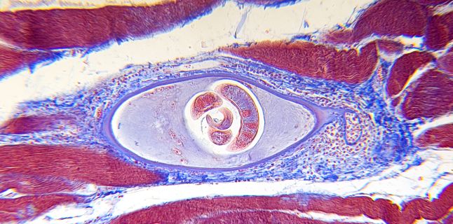
The disease is due to Trichinella spiralis larva, 1. 5-4 mm long. The development cycle takes place in the same animal organism that is infested with both adult and infected larvae. Among animals, the pig, the rat, the fox, the bear, the dog, the cat and other carnivorous animals represent the virus reservoir, from where the worm can be transmitted to humans. The man ingests meat, usually pig, infested with trichin-truncated larvae, which are released by cyst digestion by. Larvae become free and turn into adults in 5-7 days, being eliminated from the gut after the males fertilize the females that each deposit about 1500 eggs, from which new larvae develop.
They cross the intestinal wall and cross the blood, liver and lung, through the large circulation in the skeletal muscles. Here it fixes and develops and after 50 days it surrounds a cystic wall, which is formed in about 3 months. After 6 months, the calcification of the cystic wall occurs, leaving the living larva in the interior, and thus, by ingestion with the insufficiently thermally prepared meat to infect other organisms. It depends on the amount of ingested worms and on the body's reactivity. For these reasons, incubation varies from 9-28 days.
During the invasion, gastrointestinal signs appear: vague, nausea, anorexia, sometimes diarrhea. The state period begins with the larvae dissemination in various muscular territories. There is intense pain in the muscles, with local swelling. Miscellaneous moves become painful, painful (walking, chewing, swallowing, breathing). On the skin there are various rashes or scarlatiniformes, which cause itching, subangular haemorrhages.
Swelling of the periorbital region or the entire face occurs. Fever 38-39 degrees, several days. Severe forms of neuropsychiatric disorders, dizziness, mental confusion, meningitis, sometimes coma (due to brain larva infestation). Other symptoms include lung damage, cough, sputum with blood. Also, in myocardial damage are cardiac noises, tachycardia, breathing, sometimes galloping.
After 7-10 days, laboratory changes occurred: white blood cells (20,000-30,000 / mm3) with increased neutrophils and eosinophils (sometimes over 50%, less than 80%). VSH usually normal, transaminases are elevated and may appear gradually: anemia, hypoalbuminemia and hypoglycemia. The larval dissemination period lasts for 20-30 days. Convalescence coincides with larval closure and the gradual disappearance of symptoms. The most common are the nerve, given by the brain's location.
Signs are either diffuse (meningitis, encephalitis), or outbreak (depending on location, signs as for any tumor process). Myocardial, subclinical complications should be detected by careful clinical and ECG monitoring. Positive diagnosis is based on the clinical signs described, reinforced by epidemiological data - inadequately consumed meat consumption, usually the occurrence of family outbreaks or in collectivities. The lab gives precious data; . Thus, the intradermal test with 1 / 10,000 trichin antigen, which is performed with the control, rapidly gives within 5-30 minutes a whitish swelling with a diameter of about.
5 mm, surrounded by a red area. After 18-24 hours, local late reaction occurs (chronic infections). The test is positive from the 20th day of the disease to 85% of the cases and remains positive for years. The serological samples can be made: the larval (early and specific) precipitation reaction, the flocculation tests on the blade or in the tubes, the complement fixation reaction. The safest diagnostic test remains the muscular biopsy, which is performed after 3-4 weeks by harvesting from the most infected muscles and looking at the microscope or trichinoscope.
Differential diagnosis is made with meningoencephalitis, septicemia, typhoid fever, dermatomyositis, polymyositis, poliomyelitis, polyneuritis. In mild and moderate forms, the progression is good. These represent most forms of the disease. In severe forms with neurological complications and occurring on previously affected organisms, the evolution may take serious forms, the prognosis being reserved. Death can occur in 5-10% of cases.
The disease requires hygienic-dietary treatment that consists of bed rest, rich in carbohydrates and vitamins. Drug treatment is etiological, pathogenic and symptomatic. As an etiological medication, an anthelmintic is used for 5-7 days. The results are good, with general improvement. Pathogenic treatment consists of the administration of cortisone preparations with major antiallergic effect, especially in severe forms.
Symptomatic treatment aims to combat pain with analgesics and sedatives. In severe forms, glucose solutions are given in infusions, for rehydration, and tonic. It is done by the veterinary service, the disease being a zoonosis. This is done by the trichinoscopic examination of the meat given in consumption, avoiding the consumption of meat from private cuts, wild animals (wild boar). Decontamination is a measure that prevents swine infestation.
The destruction of trichinae from infected meat is by freezing, 3 months at -18grade, or rapid refrigeration at -35grade. It also uses gamma irradiation. Sufficient meat processing remains the most effective measure at all. .
Source : sfatulmedicului.ro
Views : 3867
Last Article
- Traditional
 4 effective ingredients in the fight against acne.
 - Traditional
 How to get rid of hiccups fast
 - Diets
 The wheat bran diet: the secret of lost pounds as if by magic
 - Traditional
 The recipe that will sweeten your soul this weekend!
 - I Know ?
 Is it dangerous or not to refreeze meat after thawing it?
 - Medicine
 The unusual sign of diabetes indicated by saliva.
 - I Know ?
 What to drink to boost your immune system.
 - Traditional
 10 foods that help you never age.
 - I Know ?
 What actually happens in your body if you drink a cup of coffee for breakfast
 - I Know ?
 5 surprising benefits of chia seeds
Popular Article
- (photo) Nude becomes art.
Posted: 2018-03-17, 9643 views.
- The harmful effects of air conditioning on the skin
Posted: 2017-06-08, 8333 views.
- 3 causes of dyed hair discoloration
Posted: 2017-06-15, 8203 views.
- Why early puberty occurs in girls: symptoms, favors, diagnosis and treatment
Posted: 2017-10-24, 8056 views.
- Good or bad skin treatments in the hot season
Posted: 2017-06-07, 7787 views.
Recommendations
- (photo) Nude becomes art.
Posted: 2018-03-17, 9643 views.
- The harmful effects of air conditioning on the skin
Posted: 2017-06-08, 8333 views.
- 3 causes of dyed hair discoloration
Posted: 2017-06-15, 8203 views.
- Good or bad skin treatments in the hot season
Posted: 2017-06-07, 7787 views.
- Risks of practicing sports on hot days
Posted: 2017-06-12, 7389 views.