Muscle lesions - causes, symptoms and treatments
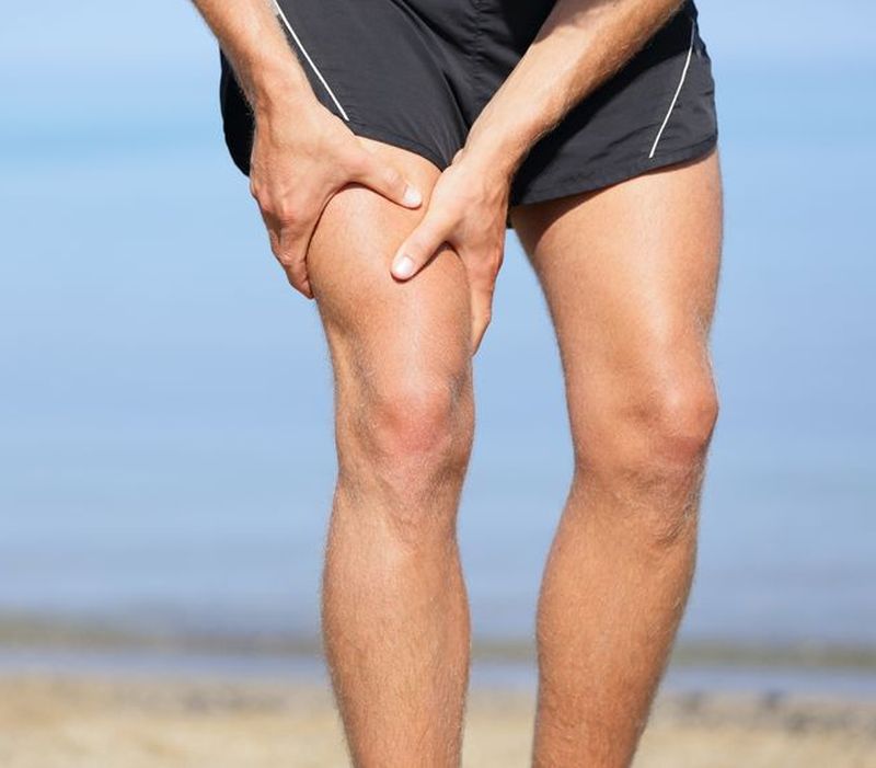
Muscle lesions are the result of trauma caused by a sudden contraction of a muscle or physical exercise at a rate that exceeds the ability of the restoring body. Doctor Omer Senol Emin, Sports Medicine Specialist, certified musculoskeletal ultrasound at Gral Medical Clinic, tells us everything we need to know about muscle damage. Table of contents Muscle lesions Muscle lesions Symptoms of muscular lesions Muscle lesions Muscle lesions Muscle lesions Muscle lesions Muscle lesions Muscle lesions Muscle lesions Muscle lesions Muscle lesions . In performance sports or amateur muscle injuries are quite high among traumas, producing in favorable conditions with predisposing, precipitating, perpetuating factors. (Muscular imbalances between agonist muscles and antagonists, eccentric contractions vs. concentric contractions), reduced mobility, kinetic chain dysfunctions and neuro - neurodegenerative traits, .
Precipitated factors: non-trained people (sedentary / athletes after a period of immobilization), undiagnosed or unrecoverable lesions (muscle cramps, inflammation, myositis, hematoma / muscle damage), non-observance of workouts or training mistakes, muscle fatigue, low muscle temperature . Perpetual Factors - repeating the same type of misdirection, inadequate restoration, inadequate hydration, non - observance of the training stages (warm - up, exercise, exertion, stretching). In the winter, for example, those who practice outdoor sports, these over-demanding conditions occur more frequently due to the low temperature of the external environment, with a more conscious warmth, the body being overburdened: once the effort itself and then the need for . Upper Symptoms Symptoms of muscle damage, sudden pain, sudden burns, burning, antalgic position (seeking and adopting a position where pain intensity decreases) and partial functional impotence of a painful point or areas when palpation occurrence of an ecchymosis or hematoma formation . This assessment, the qualification of pain, is obviously a subjective but orientative one, with the patient's response being influenced by many factors, the final diagnosis being determined by the clinical and imaging examination.
Upper Investigations and Diagnosis of Muscular Damage Muscular ultrasound (mio-entezo-articular) is the objective imaging method that confirms the muscular lesion, its type and severity, which establishes the diagnosis and therapeutic behavior. Based on previous experiences with such cases, we found that it is important to perform the first imaging examination in the first 24 hours, followed by the second ultrasound after about 48 hours, thus observing the evolution over time and the safety of a correct diagnosis. Ultrasound is an accessible, non-invasive, repeatable imaging method, and can be performed in dynamics (muscle contraction and relaxation). Option 2. The presumptive diagnosis is based on the clinical history and clinical examination.
The muscular ultrasound (mio-entezo-articular) is the objective imaging method that confirms the muscular lesion, its type and severity, which determines the therapeutic behavior. Diagnosis It is very important that diagnosis should take place as soon as possible after the onset of the trauma. The doctor will establish the diagnosis based on the anamnesis (the patient will explain the symptoms to the doctor) followed by the clinical examination. As far as diagnosis is concerned, there are other aspects that need to be taken into consideration regarding the equipment of the hurt, the experience gained by the physician with the equipment available. In the case of complex trauma involving a joint, the challenge for a physician is to evaluate the associated muscular lesions by directly intervening the doctor's experience with such cases.
Top Muscle injury treatment The treatment of muscular lesions is diverse and complex, consisting of medical and physiokinetoterapeutical treatment, and even in surgical cases. Generally, the P protocol is applied in the first 48 hours. R. I. C.
E. : P - protection - protection of the affected area R - rest - segmental maintenance I - ice - cryotherapy, or local ice applications (ice pack should not be applied directly to the skin, wrapped in towel or special gel packs . Physiotherapeutic treatment may be diverse: - Cryotherapy, Ultrasound Therapy, LASER Therapy, Dystatinic Contact Therapy (TCARE), Diadinamic Currents and Interferential Currents, TENS. Surgical treatment is usually recommended for total rupture, in these situations the patient is transported directly to the hospital. Ideally, treatment would be complex; .
THE MASS IS NOT RECOMMENDED for a minimum of 14 days. The prognosis and evolution of muscle damage are generally favorable if: the patient is immediately presenting to the doctor and observes the medical team's indications the diagnosis is made from the first moments of the trauma the medical and physiotherapeutic treatment is adequate and professionally applied, the kinetotherapeutic program starts . Following clinical and ultrasound re-evaluation, it will be decided to start the recovery, which will be done through a kinetoteptic program, prescribed by the physician and physical therapist. The moment of re-entry into sports activity will be determined by the attending physician following specific clinical examinations, testing of affected muscles (tonus and force), echographic imaging and kinetotherapeutic objectives were achieved. Throughout this process it is essential to communicate and respect the decisions of the three parties involved: sportsman, coach, doctor.
The therapeutic approach does not mean great differences between performance and amateur athletes. There are differences in the degree of supportability of the body to pain and its ability to recover, post traumatic recovery.
Source : csid.ro
Views : 3624
Last Article
- Traditional
 4 effective ingredients in the fight against acne.
 - Traditional
 How to get rid of hiccups fast
 - Diets
 The wheat bran diet: the secret of lost pounds as if by magic
 - Traditional
 The recipe that will sweeten your soul this weekend!
 - I Know ?
 Is it dangerous or not to refreeze meat after thawing it?
 - Medicine
 The unusual sign of diabetes indicated by saliva.
 - I Know ?
 What to drink to boost your immune system.
 - Traditional
 10 foods that help you never age.
 - I Know ?
 What actually happens in your body if you drink a cup of coffee for breakfast
 - I Know ?
 5 surprising benefits of chia seeds
Popular Article
- (photo) Nude becomes art.
Posted: 2018-03-17, 9749 views.
- The harmful effects of air conditioning on the skin
Posted: 2017-06-08, 8454 views.
- 3 causes of dyed hair discoloration
Posted: 2017-06-15, 8331 views.
- Why early puberty occurs in girls: symptoms, favors, diagnosis and treatment
Posted: 2017-10-24, 8178 views.
- Good or bad skin treatments in the hot season
Posted: 2017-06-07, 7908 views.
Recommendations
- (photo) Nude becomes art.
Posted: 2018-03-17, 9749 views.
- The harmful effects of air conditioning on the skin
Posted: 2017-06-08, 8454 views.
- 3 causes of dyed hair discoloration
Posted: 2017-06-15, 8331 views.
- Good or bad skin treatments in the hot season
Posted: 2017-06-07, 7908 views.
- Risks of practicing sports on hot days
Posted: 2017-06-12, 7494 views.