Tooth whitening - causes, treatment
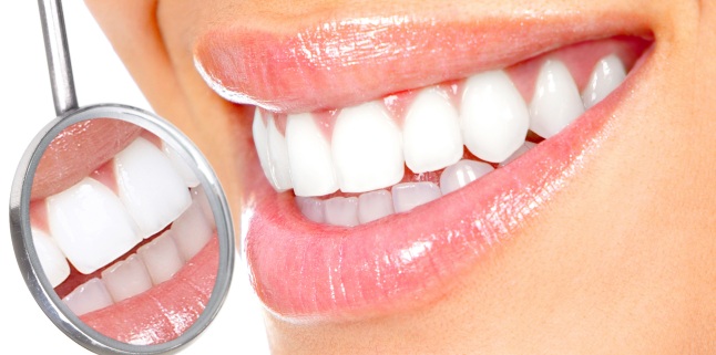
From the point of view of their mechanism of production, dystrophies can be classified as follows: A) intrinsic (endogenous) dysrhymes caused by changes in the structure or thickness of dental hard tissues (hypoplasia zones of hypermineralized areas) or the incorporation of pigments during formation . Diffusion of pigments in dental hard tissue after tooth formation (products from dental pulp necrosis, drug substances used in canal treatments), senescence or pulp bleeding may be other internal causes by which the tooth changes its color. B) extrinsic (exogenous) dysrhythmics refers to those in which external factors act on the teeth, coloring them most frequently in yellowish-brown. This can include diet pigments (chlorhexidine used in combination with food tannin gives rise to a brown-tinged color) and black or black nicotine impregnations. Sometimes, salivary cromogenic bacteria may result in the appearance of a greenish color localized to the parcel (the crown and root limit) of the teeth or the black-black, parallel to the contour line, very resistant to chemical agents. it also has an important role.
Thus, they often meet brown-black colorings on dental parcels or white-yellow deposits due to the accumulation of. Tooth color changes with simple or complicated caries lesions and color changes caused by coronal obturation materials with impregnation of dental hard dyes that appear as black-colored or determined by radicular obturation (iodoform) are other phenomena that affect the beauty of our smile. The color changes of the teeth of exogenous etiology do not alter their structure, so they are easy to remove by using abrasive pastes applied with some. Color changes caused by tetracycline Color changes occur in incisors and canines above and below tetracycline starting from IV month of intrauterine life and up to 7 years of age. The type of color change depends on the dose of the antibiotic administered (the higher it is, the greater the severity of the discoloration), the duration of the treatment (an average duration of 4-5 days is sufficient for the obvious staining of the enamel) .
Color changes caused by fluorosis Appears if in the first 7 years of life the child ingests in a concentration higher than 1 mg / liter of water. In these cases, the tooth enamel may appear colorful, marbled, moth-eyed due to fluoride interference in the calving process of the permanent teeth enamel. This causes an incomplete maturation of the enamel that becomes porous and opaque. The color of the teeth is correlated directly with the amount of absorbed fluoride and its duration. A concentration of 2-3 mg / day causes discreet changes in enamel, but also general disorders such as vomiting.
Fluoride poisoning (chronic dose of 2-3ppm) is clinically manifested by white spots, opaque and yellow or brown enamel striations of the enamel, while a concentration greater than 3 mg / day results in moderate and severe forms of fluorosis. Color is limited to enamel, it is bilateral and bimaxilar. Tooth Colors in the ElderlyIt is the most common disorder and often occurs in people over 50 years of age. During the individual's life, the teeth, which initially are bright, white, well-textured, become yellowish to brown with the passage of time. This natural phenomenon is accentuated by the consumption of tea, smoking.
Color changes due to age are ideal for the treatment of. Hemorrhagic Color Changes These disfroms occur as a result of a dental traumatic episode that occurred at an early age as a result of the penetration of the blood vessels into the dentin. These color changes do not benefit from whitening methods. Tooth whitening is done with two main substances: carbamide peroxide and. Hydrogen peroxide is, in fact, the active ingredient of bleaching systems, along with carbamide peroxide, which have a neutral pH.
Hydrogen peroxide used in the dental office has a concentration of 30-35% and has some inconveniences. It can produce dehydration and demineralisation of teeth and, being caustic, can cause soft tissue damage. It may have a negative influence on the damaged oral mucosa, so at the start of the whitening treatment, the gum tissues must be in perfect health. Bleaching occurs by penetrating peroxide into enamel and dentin and oxidizing the color spots in the tooth texture without producing any change in the intimate structure of the tooth. Whitening is initially made at the enamel level, so stains most of which are produced in dentine require a longer bleaching time and sometimes do not result.
Tooth whitening consists of applying whitening chemicals to the enamel surface. This method is known as \. 1. Office bleaching This method uses 33% hydrogen peroxide, heat and light as an aggressive method, and the dental substance may be at risk, according to some authors. It is a preferred method if a quick result is desired.
2. Home whitening (Home bleaching) Performs at home by the patient, provided it is initiated by a dentist. Carbamide peroxide is used and lasts for three or more weeks. .
Source : sfatulmedicului.ro
Views : 3077
Popular Article
- (photo) Nude becomes art.
Posted: 2018-03-17, 9591 views.
- The harmful effects of air conditioning on the skin
Posted: 2017-06-08, 8274 views.
- 3 causes of dyed hair discoloration
Posted: 2017-06-15, 8146 views.
- Why early puberty occurs in girls: symptoms, favors, diagnosis and treatment
Posted: 2017-10-24, 8003 views.
- Good or bad skin treatments in the hot season
Posted: 2017-06-07, 7740 views.
Recommendations
- (photo) Nude becomes art.
Posted: 2018-03-17, 9591 views.
- The harmful effects of air conditioning on the skin
Posted: 2017-06-08, 8274 views.
- 3 causes of dyed hair discoloration
Posted: 2017-06-15, 8146 views.
- Good or bad skin treatments in the hot season
Posted: 2017-06-07, 7740 views.
- Risks of practicing sports on hot days
Posted: 2017-06-12, 7335 views.
 4 effective ingredients in the fight against acne.
 How to get rid of hiccups fast
 The wheat bran diet: the secret of lost pounds as if by magic
 The recipe that will sweeten your soul this weekend!
  Is it dangerous or not to refreeze meat after thawing it?
  The unusual sign of diabetes indicated by saliva.
 What to drink to boost your immune system.
 10 foods that help you never age.
 What actually happens in your body if you drink a cup of coffee for breakfast
 5 surprising benefits of chia seeds