Difference: causes, symptoms, treatment

What Diphtheria is and how it is transmitted Diphtheria is an acute, transmissible infectious disease caused by Corynebacterium diphteriae, which remains at the entrance gate, multiplies and causes local phenomena (edema and false membranes) and develops a toxin that diffuses in the body, . The entrance gate is represented by the mucosal pharyngeal (96-98%), laringean (1-2%), nasal (0. 5%) - exceptionally conjunctival, genital, injured, ear - . Source of infection - the patient with a typical form (contagious before clinical onset for 10-30 days, sometimes in convalescence for up to 3 months, antibiotics shorten the period of infectivity), the atypical patient and seemingly healthy carriers (1-5% . The infectivity index varies around 20%; . The responsiveness to infection is maximum between 2 and 5 years and gradually decreases until the age of adolescence, either as a result of vaccination or as a consequence of natural infection.
Transmission path: aerogenic by drops, direct, by contact with infected or indirect persons by contaminated use, food - exceptional. What Diphtheria is and how it is transmitted How is diphtheria diagnosed Complications of diphtheria Diphtheria treatment top How to diagnose diphtheria Diphtheria toxin acts both locally and systemically. The absorbed toxin may produce myocarditis, neuritis and focal necrosis in various organs, including the kidneys, the liver and the adrenal glands. These changes are followed in a few weeks by granular degeneration of muscle fibers (sometimes with fatty degeneration of the myocardium), progressing to myolysis and finally replacing fibrous tissue destroyed with fibrous tissue. Thus, diphtheria can cause irreversible cardiac damage, and in diphtheria polyneuritis, pathological changes include regional loss of peripheral and vegetative nerve myelin tars.
Other alterations in poisoned cells are secondary to inhibition of protein synthesis. Clinical diagnosis. Incubation: 2-6 days. The clinical picture is dependent on the location and intensity of the diphtheria process. Diphtheria (pharyngitis): gradual onset with moderate fever, gradual increase, intense asthenia, nausea with or without vomiting, anorexia and pharyngeal pain.
The objective exam reveals closed-shade redness with opaline exudate that quickly turns into very solid white-pearly membranes; . They are accompanied by intense pharyngeal edema, which can be externalized. Laryngeal diphtheria (diphtheria): may be primary, as an isolated manifestation of diphtheria or secondary, by extending the pharyngeal process; . It is clinically manifested as obstructive laryngitis. Debut with fever, dysphonia, harsh cough, spasms, stridor, runny nose, dyspnea, suffocation, sometimes aphon.
The objective exam reveals false membranes on epiglottis, glottis and vocal cords, which are inflamed. The diphtheria process may extend from the larynx to the entire tracheobronchial tree, producing obstructive diphtheria tracheo-bronchitis, and the removal of false membranes as a bronchial mold. Diphtheria rhinitis: is highly contagious and is characterized by catara, nasal obstruction, monolateral submaxillary adenopathy, sometimes epistaxis, false membranes, serum-sanguinolenic secretion that can erupt narina. Skin dysthynia: usually occurs as a C infection. diphtheriae of preexisting dermatoses affecting in decreasing order of frequency, lower limbs, upper limbs, head or trunk.
Clinical aspects are similar to those of a secondary skin bacterial infection. In tropical regions, the presentation of cutaneous diphtheria occasionally includes distinct morphological appearance of Topical complications Cardiovascular: - Early toxic myocarditis occurs in the first 10 days of the disease in 25-55% of patients and is characterized by electrocardiographic abnormalities including ST and T wave changes, block variable degrees and arrhythmias, . Neurological: - Palatinate vein paralysis, occurs during the first 2 weeks with swallowing and phonation disorders (swallowing becomes difficult, nasal voice and ingested liquids may be regurgitated on the nose); . Usually polyneuria is fully healed, the time required for improvement is approximately equal to the one between exposure and the onset of symptoms. Renal: - toxic nephrosis through degenerative, haemorrhagic and necrotic lesions; .
top Diphtheria treatment Etiological: decision to administer the diphtheria antitoxin should be based on the clinical diagnosis of diphtheria without waiting for the final confirmation of the lab, each day of delay being associated with increased mortality. Antibiotherapy: its primary purpose for patients or carriers is to eradicate diphtheria bacillus and prevent transmission from patients to susceptible contacts. It is recommended to use erythromycin, penicillin G, rifampicin or clindamycin for 14 days. Destruction of diphtheria bacillus must be proven by negative cultures from samples taken within two or three consecutive days starting at least 24 hours after the end of antibiotic treatment. Supportive treatment, monitoring of respiratory and cardiac function, in the case of airway obstruction, early intubation or tracheostomy.
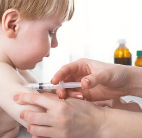
Prophylaxis is done by administering DTP (diptero-tetanopertusis). The small child is given a total of 3 doses of the vaccine, intramuscularly, at one month, followed by revaccination at 1. 5 years and at 6 years. Periodically, diphtheria toxoid may be administered after vaccination to maintain immunization. In patients with dirty plaques, tetanus toxoid tetanus vaccine is required as a DTP vaccine; .
Diphtheria infection does not necessarily give immunity, so all healed patients will then be vaccinated. They will also be evaluated and supervised for one week, all seemingly healthy individuals who have come into contact with diphtheria patients. Close contacts will be revaccinated; .
Source : csid.ro
Views : 3392
Popular Article
- (photo) Nude becomes art.
Posted: 2018-03-17, 9816 views.
- The harmful effects of air conditioning on the skin
Posted: 2017-06-08, 8529 views.
- 3 causes of dyed hair discoloration
Posted: 2017-06-15, 8410 views.
- Why early puberty occurs in girls: symptoms, favors, diagnosis and treatment
Posted: 2017-10-24, 8253 views.
- Good or bad skin treatments in the hot season
Posted: 2017-06-07, 7983 views.
Recommendations
- (photo) Nude becomes art.
Posted: 2018-03-17, 9816 views.
- The harmful effects of air conditioning on the skin
Posted: 2017-06-08, 8529 views.
- 3 causes of dyed hair discoloration
Posted: 2017-06-15, 8410 views.
- Good or bad skin treatments in the hot season
Posted: 2017-06-07, 7983 views.
- Risks of practicing sports on hot days
Posted: 2017-06-12, 7559 views.
 4 effective ingredients in the fight against acne.
 How to get rid of hiccups fast
 The wheat bran diet: the secret of lost pounds as if by magic
 The recipe that will sweeten your soul this weekend!
  Is it dangerous or not to refreeze meat after thawing it?
  The unusual sign of diabetes indicated by saliva.
 What to drink to boost your immune system.
 10 foods that help you never age.
 What actually happens in your body if you drink a cup of coffee for breakfast
 5 surprising benefits of chia seeds